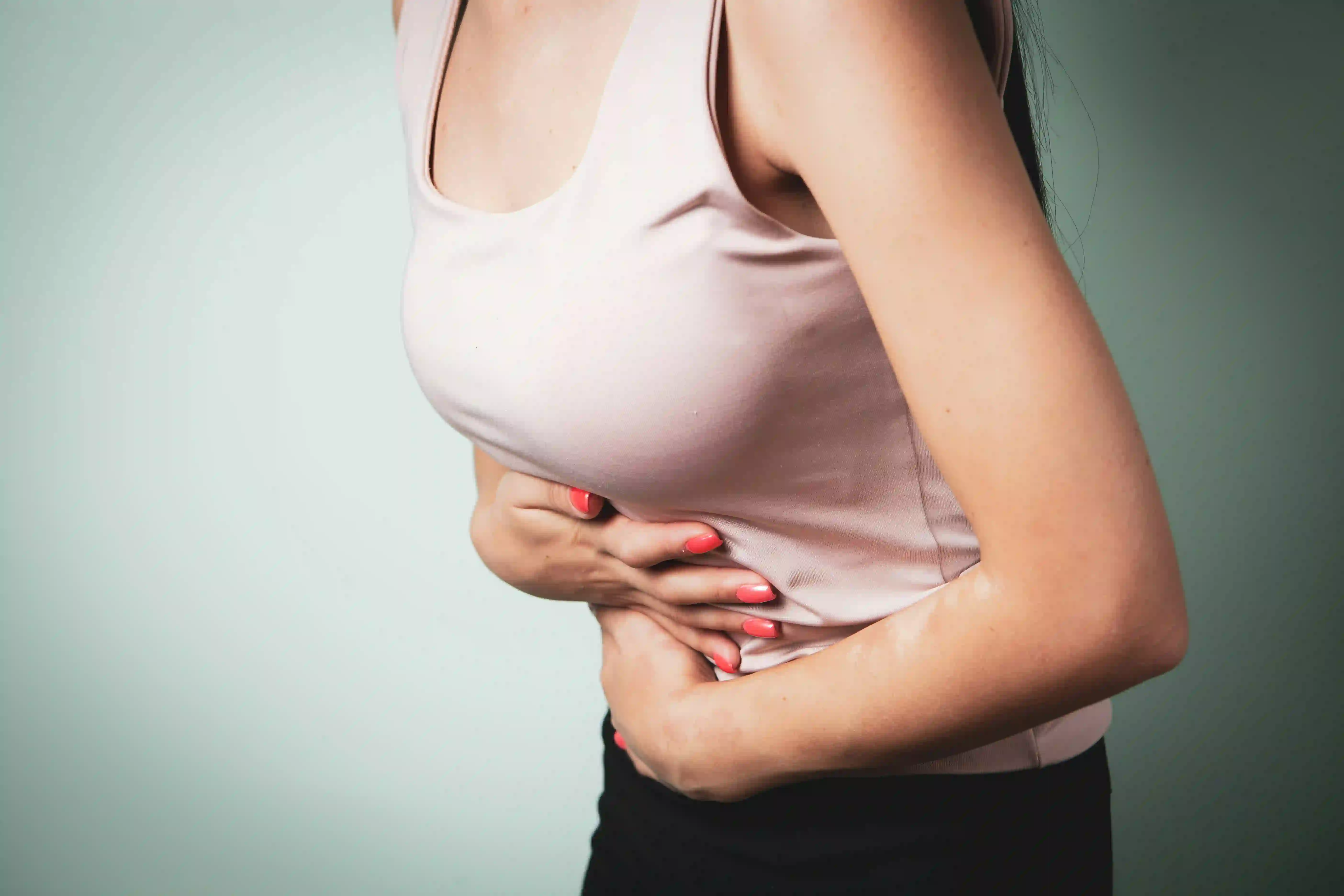
She had been soaking through pads every 45 minutes for the first three days of every period since she was 26. She kept a change of clothing at work. She booked nothing for the first week of each month because she could not predict whether she would be functional. She had been anaemic for years and took iron tablets that barely kept pace.
She was 38 when she finally described this to me. Not because she had not seen doctors before - she had. But because she had been told, each time, that some women just have heavy periods.
She had fibroids. Multiple, significant, treatable fibroids. That had been doing this to her body for over a decade while nobody thought to look properly.
Nkechi's story is not unusual. It is representative of a pattern I see repeatedly: Black and African women enduring significant fibroid symptoms for years - sometimes a decade or more - before receiving adequate investigation. Their symptoms are normalised. Their pain is attributed to character. Their bleeding is managed with iron tablets rather than investigated at source.
Up to 80% of Black women will develop uterine fibroids by the age of 50. This is not a minority experience. It is the majority one.
What fibroids are and why they grow
Fibroids are tumours of smooth muscle and fibrous connective tissue that develop in or around the uterus. They range enormously in size - from a pea to, in rare cases, the size of a large melon filling the entire abdomen. They can be solitary or multiple, submucous (inside the uterine cavity), intramural (within the uterine wall), or subserosal (on the outer surface of the uterus). Their location determines more than their size what symptoms they cause.
Fibroids are oestrogen-dependent - they grow in the presence of oestrogen and shrink after menopause when oestrogen falls. They also respond to progesterone. This is why they most commonly develop in the reproductive years, typically in the 30s and 40s, and why conditions that increase oestrogen exposure (obesity, never having been pregnant, early menarche) are associated with higher fibroid risk.
Why Black and African women are disproportionately affected
The disparity is striking and well-documented: Black women develop fibroids 2-3 times more often than white women, develop them approximately 10 years earlier, and have more severe disease at the time of diagnosis.
Genetic factors: Specific genetic variants associated with fibroid development are more common in women of African ancestry. Genome-wide association studies have identified multiple genetic loci that differ in prevalence between African-ancestry and European-ancestry women.
Vitamin D deficiency: Vitamin D appears to inhibit fibroid growth through effects on cell proliferation and inflammation. Black women living in the UK are almost universally vitamin D deficient. Several studies have found that lower vitamin D levels are associated with higher fibroid risk, and that vitamin D supplementation may reduce fibroid growth rates.
Hormonal sensitivity: There is evidence that fibroid tissue in Black women may express oestrogen and progesterone receptors differently, making it more responsive to hormonal stimulation.
Stress: Chronic psychosocial stress - and specifically the physiological effects of experiencing racism and discrimination - may promote inflammatory conditions that accelerate fibroid growth. This remains an area of active research.
Symptoms - what fibroids actually cause
Not all fibroids cause symptoms. Smaller fibroids, particularly those that are intramural or subserosal, may be incidental findings on imaging done for other reasons. When they do cause symptoms, the pattern depends significantly on location.
Heavy menstrual bleeding: The most common and most disruptive symptom. Submucous fibroids (inside the uterine cavity) cause the most severe bleeding, because they increase the surface area of the endometrium (uterine lining) that bleeds each month and disrupt the mechanisms that normally limit menstrual blood loss. Passing large clots, soaking through protection within an hour, flooding incidents, and periods lasting more than 7 days are all abnormal and warrant investigation. Chronic heavy bleeding causes iron deficiency anaemia in most women with significant fibroids.
Pelvic pain and pressure: A dull, heavy ache or feeling of fullness in the lower abdomen. Larger fibroids can cause a constant sensation of pelvic pressure. Fibroid degeneration (when a fibroid outgrows its blood supply and dies) causes acute, severe pain requiring urgent assessment.
Painful periods (dysmenorrhoea): Cramping significantly worse than baseline, often requiring strong analgesia and sometimes causing incapacitation.
Urinary symptoms: Fibroids pressing on the bladder cause urinary frequency and urgency. Fibroids pressing on the urethra can cause difficulty emptying the bladder.
Bowel symptoms: Large posterior fibroids can cause constipation or a sensation of incomplete bowel emptying.
Fertility and pregnancy: Submucous fibroids distort the uterine cavity, interfering with implantation and increasing miscarriage risk. Intramural fibroids above a certain size also affect fertility. Fibroids in pregnancy can cause pain (particularly degeneration), increase risk of preterm labour, malpresentation, and caesarean section.
Diagnosis
A standard pelvic ultrasound (transabdominal and transvaginal) is the first-line investigation and adequately identifies most fibroids. It characterises their size, number, and location.
Saline infusion sonography (SIS) or hysteroscopy provides better assessment of the uterine cavity specifically - important for planning treatment of submucous fibroids.
MRI provides the most detailed assessment for surgical planning - mapping the exact location of multiple fibroids and their relationship to the uterine cavity and blood supply.
Treatment options
Watchful waiting: Appropriate for small asymptomatic fibroids. Review annually with symptom monitoring.
Medical management of bleeding: Tranexamic acid (taken during periods) reduces menstrual blood loss by approximately 50% by inhibiting clot breakdown. Not hormonal. The levonorgestrel IUS (Mirena coil) significantly reduces menstrual blood loss in many women with fibroids, though it may be expelled if the uterine cavity is significantly distorted. GnRH agonists (leuprorelin, goserelin) induce a temporary menopause, shrinking fibroids by 30-60% and resolving bleeding. Used short-term before surgery to reduce fibroid size and improve anaemia. Relugolix/relugolix combination (Ryeqo) is a newer oral option with fewer side effects.
Myomectomy: Surgical removal of fibroids while preserving the uterus. The procedure of choice for women who wish to preserve fertility or who wish to keep their uterus. Can be performed hysteroscopically (for submucous fibroids), laparoscopically, or via open surgery depending on fibroid number, size, and location.
Hysterectomy: Surgical removal of the uterus - a definitive cure. Should not be the automatic first surgical offer. If you have been offered only hysterectomy and wish to preserve your uterus, you are entitled to ask specifically about myomectomy and whether it is feasible for your fibroid pattern.
Uterine Fibroid Embolisation (UFE): A minimally invasive radiological procedure. A catheter is inserted into the femoral artery and guided to the uterine arteries, where tiny particles are injected to block the blood supply to fibroids. Fibroids then shrink and symptoms improve significantly in approximately 85% of women. Recovery is faster than surgery. Not recommended for women planning future pregnancies. Ask your gynaecologist whether UFE is offered - not all hospitals provide it.
Sources: Stewart EA et al, Nature Reviews Disease Primers 2016; Marsh EE et al, Seminars in Reproductive Medicine 2019; NICE Clinical Guideline NG88 - Heavy Menstrual Bleeding (2018, updated 2021); Bulun SE, NEJM 2013; Wise LA et al, American Journal of Epidemiology 2011; NHS England - Uterine Fibroid Embolisation commissioning guidance.